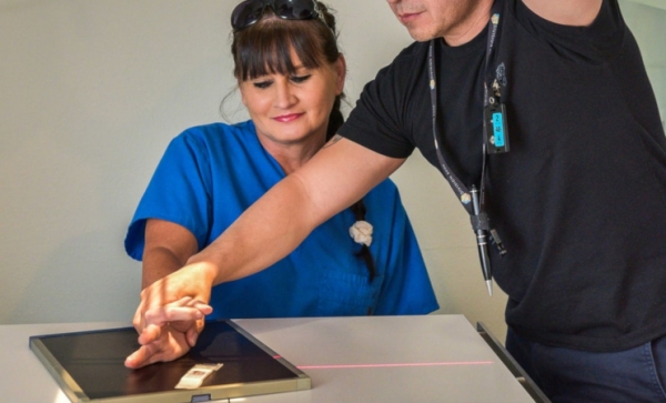
The safety of the patient is guaranteed if the recommended limit values to avoid cavitation and to overheat are observed. Let's see how the X-Ray Protection executed its early invention!
Preliminary Remark
The devices that work with alternating magnetic fields in the radio frequency range, such as magnetic resonance tomography (MRT), were not yet used. In 1973, MRT was developed as an imaging method by Paul Christian Lauterbur (1929–2007), with significant contributions from
Sir Peter Mansfield (1933–2017).
Here, there is a possibility that jewelry or piercings will become very hot; on the other hand, a high tensile force is exerted on the jewelry, which in the worst case can lead to tearing. To avoid pain and injuries, the jewelry should be removed beforehand if it is ferromagnetic.
Cardiac pacemakers, defibrillator systems, and large tattoos in the examination area containing metal-containing color pigments can heat up or cause skin burns up to degree II or cause the implants to malfunction.
Photoacoustic tomography (PAT) is a method of photoacoustic imaging that uses the photoacoustic influence and does not use ionizing radiation.
It functions in the tissue to be studied without interaction with high-speed laser pulses that produce ultrasound. The local diffusion of the light gives rise to sudden heat generation and the resulting thermal expansion.
This essentially produces acoustic broadband waves. The initial distribution of absorbed energy can be reconstructed by calculating the outgoing ultrasonic waves with suitable ultrasonic transducers.
Detection of Radiation Exposure
To better estimate radiation protection, the number of X-ray examinations, including the dose, has been recorded annually in Europe since 2007. However, no complete data from the Federal Statistical Office is available for conventional X-ray examinations.
In 2014, around 135 million x-ray examinations were estimated for Germany, including around 55 million x-ray examinations in the dental field. The mean effective dose from X-ray examinations per inhabitant in Germany for 2014 was around 1.55 mSv (around 1.7 X-ray examinations per inhabitant and year). The proportion of dental x-rays is 41% but only accounts for 0.4% of the collective effective dose.
In Germany, the X-ray Ordinance (RöV) has stipulated in Section 28 since 2002 that the attending physician has to keep X-ray passports ready and offer them to the person being examined.
Information about the patient's X-ray examinations was entered there to avoid unnecessary repeat examinations and to have the possibility of comparison with previous images. With the entry into force of the new Radiation Protection Ordinance on December 31, 2018, this obligation no longer applies. Radiation shielding became so essential to guarantee safety.
In Austria and Switzerland, X-ray passports are currently only available voluntarily. In principle, it always has a justifying indication for the use of X-rays to the patient. In connection with medical treatment, informed consent refers to the patient's consent to all interventions and other medical measures.


